


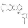
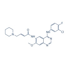
Icotinib 610798-31-7
Product Description
.cp_wz table {border-top: 1px solid #ccc;border-left:1px solid #ccc; } .cp_wz table td{border-right: 1px solid #ccc; border-bottom: 1px solid #ccc; padding: 5px 0px 0px 5px;} .cp_wz table th {border-right: 1px solid #ccc;border-bottom: 1px solid #ccc; padding: 5px 0px 0px 5px;}
Molecular Weight:
391.42 Icotinib is a potent and specific EGFR Inhibitor with IC50 of 5 nM, including the EGFR, EGFR(L858R), EGFR(L861Q), EGFR(T790M) and EGFR(T790M, L858R).
Biological Activity
Icotinib inhibits EGFR activity in a dose-dependent manner, with an IC50
value of 5 nM and complete inhibition at 62.5 nM. Icotinib selectively
solely inhibits the EGFR members including the wild type and mutants
with inhibition efficacies of 61-99%. Icotinib blocks EGFR-mediated
intracellular tyrosine phosphorylation in human epidermoid carcinoma
A431 cells in a dose-dependent manner.
Meanwhile, in our proliferation
assay performed on A431, BGC-823, A549, H460, HCT8, KB and Bel-7402 cell
lines, we found that the relative sensitivity of cell lines to Icotinib
is A431 > BGC-823 > A549 > H460 > KB > HCT8 and
Bel-7402. Icotinib exhibits a broad spectrum of antitumor activity and
it is especially effective against tumors expressing higher levels of
EGFR.
Icotinib
shows an antitumor effect in different types of xenografts. Icotinib
inhibits tumor growth at a rate of 51.5%, 31.0% and 67.4% in the A431,
A549 and H460 xenografts at a dose of 120 mg/kg, respectively.
Biochemical kinase assays
In the in vitro kinase assays, 2.4 ng/μL EGFR protein is mixed with 32
ng/μL Crk in 25 μL kinase reaction buffer containing 1 μM cold ATP and 1
μCi 32P-γ-ATP. The mix is incubated with Icotinib at 0, 0.5,
2.5, 12.5 or 62.5 nM on ice for 10 min followed by incubation at 30 °C
for 20 min. After quenching with SDS sample buffer at 100 °C for 4 min,
the protein mix is resolved by electrophoresis in a 10% SDS-PAGE gel.
The dried gel is then exposed to the PhosphorImager to detect
radioactivity. Quantification is performed by ImageQuant software. In
this methodology the radioactive signal inversely correlates with kinase
activity.
Method
Cells (103 /well) are seeded into 96-well plates in RPMI-1640 medium containing 10% FBS and grown in a 5% CO2 incubator at 37 °C. After 24 h, cells are treated with Icotinib at 0,
0.78, 1.56, 3.125, 6.25, 12.5 or 25 μM for 96 h. Cell proliferation is
calculated by subtracting the mean absorbance value on day 0 from the
mean absorbance value on day 4.
Contact us if you need more details on 610798-31-7. We are ready to answer your questions on packaging, logistics, certification or any other aspects about Icotinib 610798-31-7、610798-31-7 Icotinib. If these products fail to match your need, please contact us and we would like to provide relevant information.
Product Categories : Protein Tyrosine Kinase > EGFR Inhibitor
Other Products
Hot Products
Astragaloside AChlortetracycline HCl 64-72-2Paclitaxel 33069-62-4Dexamethasone Acetate 1177-87-3Dinaciclib (SCH727965) 779353-01-4CHIR-124 405168-58-3Ro3280 1062243-51-9TAME 901-47-3CCG-1423 285986-88-110058-F4 403811-55-2Dabigatran (BIBR 953) 211914-51-1H 89 2HCl 130964-39-5T0901317 293754-55-9Aprepitant 170729-80-3Turofexorate Isopropyl (XL335) 629664-81-9BMS-378806 357263-13-9