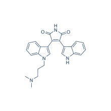
Staurosporine
Product Description
.cp_wz table {border-top: 1px solid #ccc;border-left:1px solid #ccc; } .cp_wz table td{border-right: 1px solid #ccc; border-bottom: 1px solid #ccc; padding: 5px 0px 0px 5px;} .cp_wz table th {border-right: 1px solid #ccc;border-bottom: 1px solid #ccc; padding: 5px 0px 0px 5px;}
Molecular Weight:
466.53 Staurosporine is a potent PKC Inhibitor for PKCα, PKCγ and PKCη with IC50 of 2 nM, 5 nM and 4 nM, less potent to PKCδ (20 nM), PKCε (73 nM) and little active to PKCζ (1086 nM). Phase 3.
Biological Activity
Protocol(Only for Reference)
Kinase Assay:
[1]
Cell Assay:
[3]
Animal Study:
[8]
Conversion of different model animals based on BSA (Value based on data from FDA Draft Guidelines)
For example, to modify the dose of resveratrol used for a mouse (22.4 mg/kg) to a dose based on the BSA for a rat, multiply 22.4 mg/kg by the Km factor for a mouse and then divide by the Km factor for a rat. This calculation results in a rat equivalent dose for resveratrol of 11.2 mg/kg.
Clinical Trial Information( data from http://clinicaltrials.gov, updated on 2015-02-07)
Chemical Information
Molarity Calculator
Dilution Calculator
Molecular Weight Calculator
Research Area
Customer Reviews (3)
Molecular Weight:
466.53 Staurosporine is a potent PKC Inhibitor for PKCα, PKCγ and PKCη with IC50 of 2 nM, 5 nM and 4 nM, less potent to PKCδ (20 nM), PKCε (73 nM) and little active to PKCζ (1086 nM). Phase 3.
Biological Activity
| Description | Staurosporine is a potent PKC inhibitor for PKCα, PKCγ and PKCη with IC50 of 2 nM, 5 nM and 4 nM, less potent to PKCδ (20 nM), PKCε (73 nM) and little active to PKCζ (1086 nM). Phase 3. | |||||
|---|---|---|---|---|---|---|
| Targets | PKCα [1] | PKCη [1] | PKCγ [1] | PKCδ [1] | PKCε [1] | PKCζ [1] |
| IC50 | 2 nM | 4 nM | 5 nM | 20 nM | 73 nM | 1086 nM |
| In vitro | Staurosporine, a microbial alkaloid, significantly inhibits protein kinase C from rat brain with IC50 of 2.7 nM. Staurosporine displays strong inhibitory effect against HeLa S3 cells with IC50 of 4 nM. [1] Staurosporine also inhibits a variety of other protein kinases, including PKA, PKG, phosphorylase kinase, S6 kinase, Myosin light chain kinase (MLCK), CAM PKII, cdc2, v-Src, Lyn, c-Fgr, and Syk with IC50 of 15 nM, 18 nM, 3 nM, 5 nM, 21 nM, 20 nM, 9 nM, 6 nM, 20 nM, 2 nM, and 16 nM, respectively. Staurosporine (1 μM) induces >90% Apoptosis in PC12 cells. Consistently, Staurosporine treatment induces a rapid and prolonged elevation of intracellular free calcium levels [Ca2+]i, accumulation of mitochondrial reactive oxygen species (ROS), and subsequent mitochondrial dysfunction. The apoptosis of MCF7 cells induced by Staurosporine can be enhanced by the expression of functional caspase-3 via caspase-8 activation and Bid cleavage. Staurosporine treatment at 1 μM only partially inhibits IL-3-stimulated Bcl2 phosphorylation but completely blocks PKC-mediated Bcl2 phosphorylation. Staurosporine induces apoptosis of human foreskin fibroblasts AG-1518, depending on the lysosomal cathepsins D mediated cytochrome c release and caspase activation. In addition to activating the classical mitochondrial apoptosis pathway, Staurosporine triggers a novel intrinsic apoptosis pathway, relying on the activation of caspase-9 in the absence of Apaf-1. | |||||
| In vivo | In the gerbil and rat ischemia models, Staurosporine pretreatment (0.1-10 ng) before ischemia prevents neuronal damage in a dose-dependent manner, suggesting the involvement of PKC in CAl pyramidal cell death after ischemia. | |||||
| Features | | |||||
Protocol(Only for Reference)
Kinase Assay:
[1]
| Enzyme assay and binding assay | Protein kinase C is assayed in a reaction mixture (0.25 mL) containing 5 μmol of Tris/HCl, pH 7.5, 2.5 μmol of magnesium acetate, 50 μg of histone II S, 20 μg of phosphatidylserine, 0.88 μg of diolein, 125 nmol of CaCl2, 1.25 nmol of [γ-32]ATP (5-10 × 104 cpm/nmol) and 5 μg of partially purified enzyme. The binding of [3H]PDBu to protein kinase C is determined: Reaction mixture (200 μL contained 4 μmo1 of Tris/malate, pH 6.8, 20 μmol of KCl, 30 nmol of CaC12, 20 μg of phosphatidylserine, 5 μg of partially purified protein kinase C, 0.5% (final concentration) of DMSO,10 pmol of [3H]PDBu (l-3 × 10⁴ cpm/pmol) and 10 μL of various amounts of Staurosporine. |
|---|
Cell Assay:
[3]
| Cell lines | PC12 |
|---|---|
| Concentrations | Dissolved in DMSO, final concentration 1 μM |
| Incubation Time | ~32 hours |
| Method | Cells are exposed to Staurosporine for ~32 hours. Cells are fixed in 4% paraformaldehyde and stained with the DNA-binding dye Hoechst 33342. Cells are visualized under epifluorescence illumination, and the percentage of apoptotic cells (cells with condensed and fragmented DNA) is determined. |
Animal Study:
[8]
| Animal Models | Male Mongolian gerbils or male Wistar rats subjected to transient ischemia |
|---|---|
| Formulation | Dissolved in DMSO, and diluted in saline |
| Dosages | ~10 ng |
| Administration | Stereotaxically administered into the bilateral CAl subfield of the hippocampus |
Conversion of different model animals based on BSA (Value based on data from FDA Draft Guidelines)
| Species | Baboon | Dog | Monkey | Rabbit | Guinea pig | Rat | Hamster | Mouse |
| Weight (kg) | 12 | 10 | 3 | 1.8 | 0.4 | 0.15 | 0.08 | 0.02 |
| Body Surface Area (m2) | 0.6 | 0.5 | 0.24 | 0.15 | 0.05 | 0.025 | 0.02 | 0.007 |
| Km factor | 20 | 20 | 12 | 12 | 8 | 6 | 5 | 3 |
| Animal A (mg/kg) = Animal B (mg/kg) multiplied by | Animal B Km |
| Animal A Km |
For example, to modify the dose of resveratrol used for a mouse (22.4 mg/kg) to a dose based on the BSA for a rat, multiply 22.4 mg/kg by the Km factor for a mouse and then divide by the Km factor for a rat. This calculation results in a rat equivalent dose for resveratrol of 11.2 mg/kg.
| Rat dose (mg/kg) = mouse dose (22.4 mg/kg) × | mouse Km(3) | = 11.2 mg/kg |
| rat Km(6) |
Clinical Trial Information( data from http://clinicaltrials.gov, updated on 2015-02-07)
| NCT Number | Recruitment | Conditions | Sponsor /Collaborators | Start Date | Phases |
|---|---|---|---|---|---|
| NCT00301938 | Completed | Accelerated Phase Chronic Myelogenous Leukemia|Adult Acute Megakaryoblastic Leukemia (M7)|Adult Acute Minimally Differentiated Myeloid Leukemia (M0 ...more Accelerated Phase Chronic Myelogenous Leukemia|Adult Acute Megakaryoblastic Leukemia (M7)|Adult Acute Minimally Differentiated Myeloid Leukemia (M0)|Adult Acute Monoblastic Leukemia (M5a)|Adult Acute Monocytic Leukemia (M5b)|Adult Acute Myeloblastic Leukemia With Maturation (M2)|Adult Acute Myeloblastic Leukemia Without Maturation (M1)|Adult Acute Myeloid Leukemia With 11q23 (MLL) Abnormalities|Adult Acute Myeloid Leukemia With Inv(16)(p13;q22)|Adult Acute Myeloid Leukemia With t(15;17)(q22;q12)|Adult Acute Myeloid Leukemia With t(16;16)(p13;q22)|Adult Acute Myeloid Leukemia With t(8;21)(q22;q22)|Adult Acute Myelomonocytic Leukemia (M4)|Adult Acute Promyelocytic Leukemia (M3)|Adult Erythroleukemia (M6a)|Adult Pure Erythroid Leukemia (M6b)|Blastic Phase Chronic Myelogenous Leukemia|Myelodysplastic/Myeloproliferative Neoplasms|Previously Treated Myelodysplastic Syndromes|Recurrent Adult Acute Lymphoblastic Leukemia|Recurrent Adult Acute Myeloid Leukemia|Relapsing Chronic Myelogenous Leukemia|Secondary Acute Myeloid Leukemia|T-cell Adult Acute Lymphoblastic Leukemia|Untreated Adult Acute Lymphoblastic Leukemia|Untreated Adult Acute Myeloid Leukemia | National Cancer Institute (NCI) | December 2005 | Phase 1 |
| NCT00301938 | Completed | Accelerated Phase Chronic Myelogenous Leukemia|Adult Acute Megakaryoblastic Leukemia (M7)|Adult Acute Minimally Differentiated Myeloid Leukemia (M0 ...more Accelerated Phase Chronic Myelogenous Leukemia|Adult Acute Megakaryoblastic Leukemia (M7)|Adult Acute Minimally Differentiated Myeloid Leukemia (M0)|Adult Acute Monoblastic Leukemia (M5a)|Adult Acute Monocytic Leukemia (M5b)|Adult Acute Myeloblastic Leukemia With Maturation (M2)|Adult Acute Myeloblastic Leukemia Without Maturation (M1)|Adult Acute Myeloid Leukemia With 11q23 (MLL) Abnormalities|Adult Acute Myeloid Leukemia With Inv(16)(p13;q22)|Adult Acute Myeloid Leukemia With t(15;17)(q22;q12)|Adult Acute Myeloid Leukemia With t(16;16)(p13;q22)|Adult Acute Myeloid Leukemia With t(8;21)(q22;q22)|Adult Acute Myelomonocytic Leukemia (M4)|Adult Acute Promyelocytic Leukemia (M3)|Adult Erythroleukemia (M6a)|Adult Pure Erythroid Leukemia (M6b)|Blastic Phase Chronic Myelogenous Leukemia|Myelodysplastic/Myeloproliferative Neoplasms|Previously Treated Myelodysplastic Syndromes|Recurrent Adult Acute Lymphoblastic Leukemia|Recurrent Adult Acute Myeloid Leukemia|Relapsing Chronic Myelogenous Leukemia|Secondary Acute Myeloid Leukemia|T-cell Adult Acute Lymphoblastic Leukemia|Untreated Adult Acute Lymphoblastic Leukemia|Untreated Adult Acute Myeloid Leukemia | National Cancer Institute (NCI) | December 2005 | Phase 1 |
| NCT00098956 | Completed | Extensive Stage Small Cell Lung Cancer|Recurrent Small Cell Lung Cancer | National Cancer Institute (NCI) | January 2005 | Phase 2 |
| NCT00098956 | Completed | Extensive Stage Small Cell Lung Cancer|Recurrent Small Cell Lung Cancer | National Cancer Institute (NCI) | January 2005 | Phase 2 |
| NCT00082017 | Completed | Lymphoma, Large-Cell, Ki-1|Lymphoma, T-Cell | National Cancer Institute (NCI)|National Institutes of He ...more National Cancer Institute (NCI)|National Institutes of Health Clinical Center (CC) | April 2004 | Phase 2 |
Chemical Information
| Molecular Weight (MW) | 466.53 |
|---|---|
| Formula | C28H26N4O3 |
| CAS No. | 62996-74-1 |
| Storage | 3 years -20℃Powder |
|---|---|
| 6 months-80℃in solvent (DMSO, water, etc.) | |
| Synonyms | CGP 41251 |
| Solubility (25°C) * | In vitro | DMSO | 4 mg/mL (8.57 mM) |
|---|---|---|---|
| Water | <1 mg/mL ( | ||
| Ethanol | <1 mg/mL ( | ||
| * <1 mg/ml means slightly soluble or insoluble. * Please note that Selleck tests the solubility of all compounds in-house, and the actual solubility may differ slightly from published values. This is normal and is due to slight batch-to-batch variations. | |||
| Chemical Name | 9,13-Epoxy-1H,9H-diindolo[1,2,3-gh:3',2',1'-lm]pyrrolo[3,4-j][1,7]benzodiazonin-1-one, 2,3,10,11,12,13-hexahydro-10-methoxy-9-methyl-11-(methylamino)-, [9S-(9α,10β,11β,13α)]- |
|---|
Molarity Calculator
Dilution Calculator
Molecular Weight Calculator
Research Area
Customer Reviews (3)
| Click to enlarge
Rating Source PLoS One 2012 7(6), e39679. Staurosporine purchased from Selleck Method Western blot Cell Lines HeLa cells Concentrations 100 nM Incubation Time 2 h Results It examined the effect on HSF1-S326 phosphorylation of drugs that have been shown to inhibit mTOR activity, including rapamycin, KUOO6379 and staurosporine. Heat shock led to phosphorylation of HSF1 on S326. Staurosporine inhibited the stress-induced activation of HSF1-S326 phosphorylation while abolishing the phosphorylation on T389 of ribosomal S6 kinase, a well-characterized substrate for mTOR. Inhibition of HSF1 phosphorylation on S326 by rapamycin suggests that this site in HSF1 is a target for the TORC1 complex.
|


